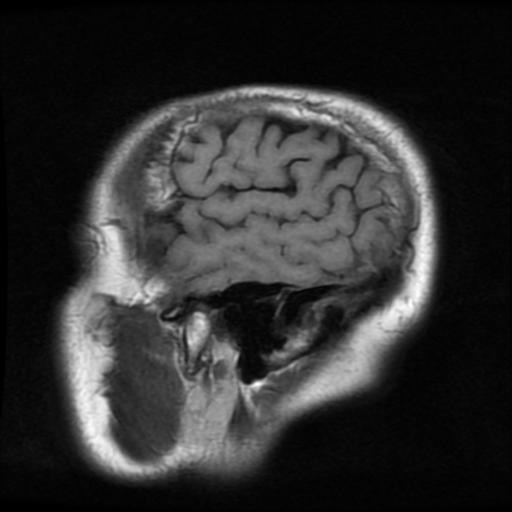
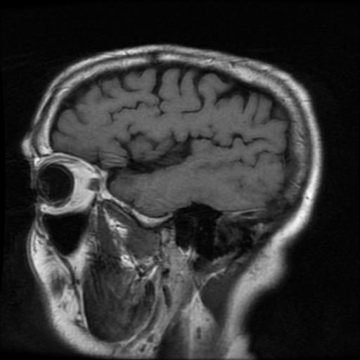
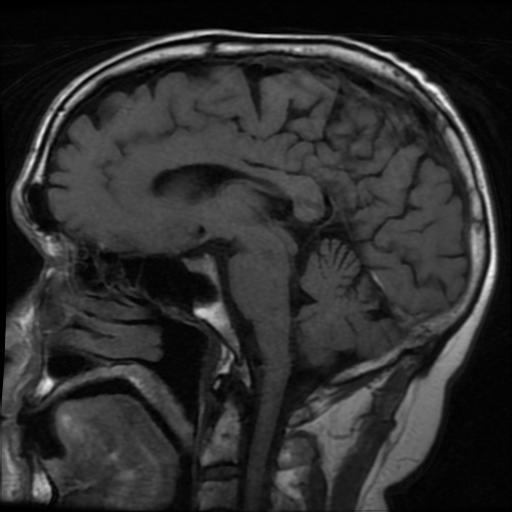
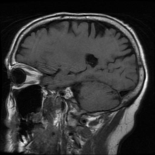
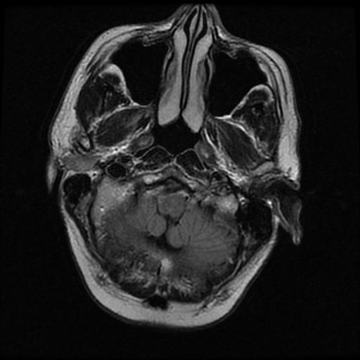
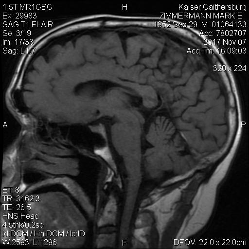
On 2017-11-07, for an hour or so, the spins of some protons in the hydrogen atoms in my brain were aligned, then tickled to make them flip. Magnetic field sensors picked up the weak signals, accumulated them, and made pictures. Here are a few samples. What do they mean? Who knows? Nothing bad, most likely. "No Worries, Mate!" as the Aussies often say.
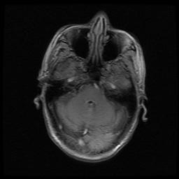 | 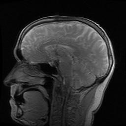 | 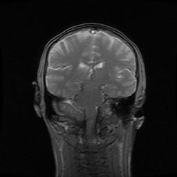 |
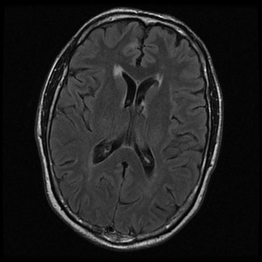
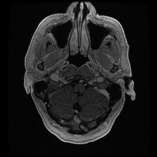
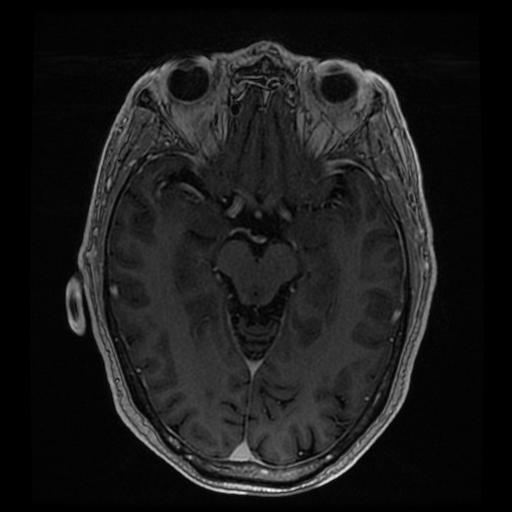
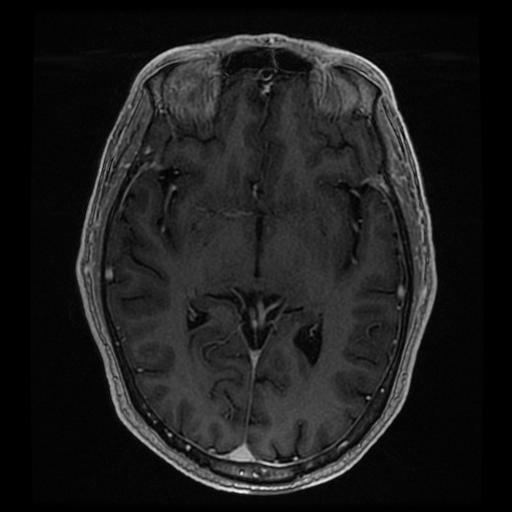
The MRI was requested after a hearing test picked up some asymmetric loss of sensitivity. Probably a random fluctuation in responses during the exam. Spending some long blocks of time holding still in the midst of noisy machinery was actually fun. And the resulting images are fascinating. A sampling:
 |  |
 |  |
 |  |
 |  |
 |  |
The physician-examiner's "findings":
No mass or abnormal enhancement is seen in the cerebellopontine angle cisterns or internal auditory canals. The 7th and 8th cranial nerve complexes appear within normal limits bilaterally.
Ventricles, sulci, and cisterns are age-appropriate in size and configuration. Midline structures including the corpus callosum, pituitary gland, and craniocervical junction are normal. There is no restricted diffusion to suggest acute infarction. Scattered T2/FLAIR hyperintense foci in the periventricular and subcortical white matter, nonspecific, likely representing mild chronic small vessel ischemic changes. A few lacunar infarcts noted in the bilateral anterior periventricular white matter. Prominent perivascular spaces. No mass lesion is identified. No abnormal parenchymal, leptomeningeal or ependymal enhancement following the administration of contrast. Major intracranial flow voids and dural venous sinuses are maintained. Visualized orbits and sinuses are normal. The mastoid air cells are clear. Large arachnoid granulation protruding into the left lateral aspect of the occipital bone.
^z - 2018-01-13